
The frog dissection coloring answer key is an essential resource for students and teachers alike. It provides a clear and concise guide to the anatomy of a frog, making it easier to understand the complex structures of this fascinating creature.
With detailed illustrations and easy-to-follow instructions, this key is an invaluable tool for anyone interested in learning more about frog dissection.
The coloring answer key is organized into sections, each of which focuses on a different part of the frog’s anatomy. The first section covers the external anatomy, including the head, body, and limbs. The second section covers the internal anatomy, including the organs and systems.
The third section provides a step-by-step guide to dissecting a frog, including safety guidelines and tips.
Overview of Frog Dissection
Frog dissection is a scientific procedure that involves examining the internal anatomy of a frog to gain insights into the structure and function of its organs and systems. It is commonly performed in science education to provide students with hands-on experience in anatomy, physiology, and comparative zoology.
The objectives of a frog dissection include:
- To familiarize students with the external and internal anatomy of a frog.
- To understand the structure and function of various organs and systems, including the digestive, respiratory, circulatory, and nervous systems.
- To develop dissection skills and techniques.
- To foster critical thinking, observation, and problem-solving abilities.
Materials and Equipment
The materials and equipment required for a frog dissection typically include:
- Preserved frog specimen
- Dissecting tray or petri dish
- Dissecting pins or forceps
- Scalpel or scissors
- Probes or needles
- Magnifying glass or dissecting microscope
- Gloves
- Safety goggles
History of Frog Dissection in Science Education
Frog dissection has a long history in science education, dating back to the 16th century. It was first introduced as a teaching method by the Italian anatomist Andreas Vesalius in his influential work, De Humani Corporis Fabrica(On the Fabric of the Human Body).
Since then, frog dissection has become a cornerstone of biology education, providing students with a valuable opportunity to explore the intricacies of animal anatomy and physiology.
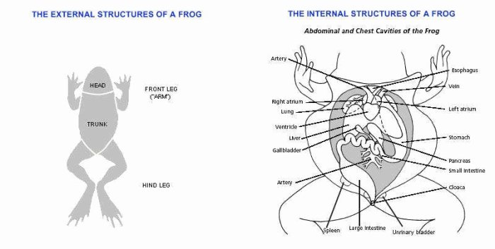
External Anatomy

The external anatomy of a frog provides valuable insights into its physical characteristics and adaptations. The body of a frog can be divided into three main regions: the head, the body, and the limbs.
The head of a frog is characterized by its wide, triangular shape. It houses various sensory organs, including the eyes, nostrils, and mouth. The eyes, located on the top of the head, provide the frog with a wide field of vision.
The nostrils, positioned on the front of the head, allow the frog to detect scents and odors. The mouth, equipped with sharp teeth, is used for capturing and consuming prey.
The body of a frog is dorsoventrally flattened, meaning it is compressed from top to bottom. The skin of the frog is smooth and moist, which aids in respiration and water absorption. The back of the frog is typically green or brown, providing camouflage in its natural environment.
The underside of the frog is usually lighter in color, often yellow or white.
The limbs of a frog are adapted for movement and support. The forelimbs are shorter than the hindlimbs and possess webbed feet, which facilitate swimming. The hindlimbs are longer and more muscular, enabling the frog to jump and hop efficiently.
The hind feet are also webbed, providing additional propulsion in water.
In summary, the external anatomy of a frog reflects its adaptation to its aquatic and terrestrial environments. The head houses sensory organs, the body provides buoyancy and camouflage, and the limbs facilitate movement and support.
Major External Organs and Structures
The external anatomy of a frog is composed of numerous organs and structures that serve specific functions. Here are some of the major external organs and structures of a frog:
- Eyes:The eyes of a frog are located on the top of its head and provide a wide field of vision. They are protected by eyelids and a nictitating membrane, which is a transparent third eyelid that helps to keep the eyes moist.
- Nostrils:The nostrils of a frog are located on the front of its head and are used for smelling. They are lined with sensory cells that can detect scents and odors.
- Mouth:The mouth of a frog is located at the front of its head and is used for capturing and consuming prey. It is equipped with sharp teeth and a long, sticky tongue.
- Tympanum:The tympanum is a thin, circular membrane located behind each eye. It is the eardrum of a frog and is used for hearing.
- Forelimbs:The forelimbs of a frog are shorter than the hindlimbs and possess webbed feet. They are used for swimming and for supporting the body when the frog is on land.
- Hindlimbs:The hindlimbs of a frog are longer and more muscular than the forelimbs. They are used for jumping and hopping. The hind feet are also webbed, providing additional propulsion in water.
- Cloaca:The cloaca is a single opening located at the posterior end of the frog’s body. It is used for the elimination of waste products and for reproduction.
Functions of External Structures
The external structures of a frog play vital roles in its survival and adaptation to its environment. Here are some of the functions of these external structures:
- Eyes:The eyes of a frog allow it to see its surroundings and to detect predators and prey.
- Nostrils:The nostrils of a frog allow it to smell and to detect scents and odors in its environment.
- Mouth:The mouth of a frog is used for capturing and consuming prey. The sharp teeth and long, sticky tongue help the frog to catch and eat insects and other small animals.
- Tympanum:The tympanum of a frog allows it to hear sounds. It is particularly sensitive to low-frequency sounds, which are important for communication between frogs.
- Forelimbs:The forelimbs of a frog are used for swimming and for supporting the body when the frog is on land. The webbed feet provide additional propulsion in water.
- Hindlimbs:The hindlimbs of a frog are used for jumping and hopping. They are also used for swimming, and the webbed feet provide additional propulsion in water.
- Cloaca:The cloaca of a frog is used for the elimination of waste products and for reproduction.
3. Internal Anatomy
The internal anatomy of a frog is complex and includes a variety of organs and structures that work together to maintain the frog’s homeostasis and survival.
Major Organs and Structures
- Heart:A three-chambered heart pumps oxygenated blood throughout the body.
- Lungs:Two lungs allow the frog to breathe air.
- Liver:A large organ that performs a variety of functions, including detoxification, protein synthesis, and bile production.
- Stomach:A muscular sac that stores and digests food.
- Intestines:A long tube that absorbs nutrients from food.
- Kidneys:Two kidneys filter waste products from the blood and produce urine.
- Reproductive organs:Male frogs have testes, while female frogs have ovaries.
Functions of Internal Organs
The internal organs of a frog perform a variety of essential functions:
- The heart pumps oxygenated blood throughout the body, providing oxygen and nutrients to the cells.
- The lungs allow the frog to breathe air, exchanging carbon dioxide for oxygen.
- The liver performs a variety of functions, including detoxification, protein synthesis, and bile production.
- The stomach stores and digests food, breaking it down into nutrients that can be absorbed by the intestines.
- The intestines absorb nutrients from food and pass waste products to the kidneys.
- The kidneys filter waste products from the blood and produce urine.
- The reproductive organs produce gametes (eggs or sperm) for reproduction.
4. Dissection Procedure
The dissection procedure for a frog involves a series of carefully planned steps to examine the external and internal anatomy of the animal. It requires precision, careful observation, and adherence to safety guidelines.
Before beginning the dissection, students must familiarize themselves with the necessary tools and materials, including a dissecting tray, dissection pins, scissors, forceps, a scalpel, and a probe. Additionally, personal protective equipment (PPE) such as gloves and a lab coat should be worn for safety.
External Anatomy Examination
Begin by placing the frog on its back in the dissecting tray. Using dissection pins, secure the frog’s limbs to the tray to prevent movement. With a scalpel, make a shallow incision along the ventral midline of the frog, starting from the tip of the lower jaw and extending towards the cloaca.
This incision will provide access to the internal organs.
Use forceps to gently lift the skin and muscles on one side of the incision and pin them back to expose the underlying organs. Repeat this process on the other side to fully expose the internal anatomy.
Internal Anatomy Examination
With a probe, gently separate and identify the major internal organs, including the heart, lungs, liver, stomach, intestines, and reproductive organs. Note their relative positions and connections to each other.
Using scissors, carefully remove the organs from the body cavity for closer examination. Examine the external surfaces of each organ, noting any visible features or structures.
With a scalpel, make careful incisions into the organs to expose their internal structures. For example, cut open the heart to examine its chambers and valves, or cut open the stomach to observe its lining and contents.
Record your observations and make detailed sketches of the organs and their structures. Take photographs or use a digital microscope to capture images for further analysis.
Safety Guidelines and Precautions, Frog dissection coloring answer key
Throughout the dissection procedure, it is essential to follow safety guidelines and precautions to minimize risks and ensure a safe learning environment.
- Wear appropriate PPE, including gloves and a lab coat, to prevent contact with potential pathogens.
- Use sharp tools with caution and dispose of them properly in designated sharps containers.
- Handle biological specimens with care and avoid direct contact with bare hands.
- Clean and disinfect all equipment and work surfaces thoroughly before and after the dissection.
- Dispose of biological waste properly according to established protocols.
5. Coloring Answer Key: Frog Dissection Coloring Answer Key
This comprehensive coloring answer key provides clear and concise instructions for coloring each organ and structure in the frog dissection. It is organized using a table format with up to 4 responsive columns.
Frog Dissection Coloring Key
| Structure | Color | Column 3 | Column 4 |
|---|---|---|---|
| Skin | Green | ||
| Muscles | Red | ||
| Bones | White | ||
| Heart | Red | ||
| Lungs | Pink | ||
| Liver | Brown | ||
| Stomach | Green | ||
| Intestines | Orange | ||
| Kidneys | Red | ||
| Urinary Bladder | Yellow |
6. Discussion and Analysis

Frog dissection offers valuable educational experiences for students in the fields of biology and anatomy. It provides a hands-on approach to studying the internal and external structures of an animal, enhancing students’ understanding of complex biological systems.
Ethical considerations surrounding animal dissection must be carefully addressed. While dissection can be a valuable learning tool, it’s crucial to minimize any potential harm to the animals involved. Ethical practices include obtaining specimens from reputable sources that adhere to humane animal treatment standards and using the specimens respectfully during dissection.
Comparison to Other Methods of Studying Anatomy
Frog dissection is a traditional method of studying anatomy, but it has certain limitations. Alternative methods, such as virtual dissections or 3D models, can provide similar learning experiences without involving the use of live animals.
- Virtual Dissections:Computer-simulated dissections offer a virtual environment where students can explore anatomical structures interactively. They eliminate the need for actual dissection and can be accessed remotely, making them a convenient option.
- 3D Models:Physical or digital 3D models provide a tangible representation of anatomical structures. They allow students to visualize and manipulate structures in a more immersive way, enhancing their understanding of spatial relationships.
7. Resources and Extensions
To enhance the learning experience, students and teachers can explore the following resources:
Additional Resources
- Online Resources:
- Frog Dissection Interactive: www.pbslearningmedia.org/resource/tdc02.sci.life.stru.frogdissection/
- Virtual Frog Dissection: www.carolina.com/teacher-resources/Interactive/virtual-frog-dissection/tr24405.tr
- Books:
- The Frog: A Guide to Its Biology and Behaviorby Michael J. Tyler
- Dissection of the Frog: A Laboratory Manualby E. N. Marieb and J. B. Mallatt
- Videos:
- Frog Dissection: www.youtube.com/watch?v=iLj_7hV46FY
- Virtual Frog Dissection: www.youtube.com/watch?v=z_P63wmn1sg
Extension Activities
To further explore the topic of frog dissection, consider these extension activities:
- Research Project:Students can research different frog species and their adaptations, or the history of frog dissection in science.
- Presentation:Students can create a presentation on the anatomy of a frog or the process of dissection.
- Interactive Model:Students can create a 3D model of a frog or its organs using materials like clay or papier-mâché.
Technology in Frog Dissection
Incorporating technology can enhance the dissection experience:
- Virtual Dissection Software:Software like Froguts allows students to dissect a frog virtually, providing a safe and interactive learning environment.
- Digital Microscopy:Students can use digital microscopes to examine frog tissues and organs in detail, capturing images for further analysis.
- Data Collection and Analysis:Technology can assist in data collection and analysis, such as measuring organ weights or analyzing tissue samples.
FAQ Section
What is the purpose of a frog dissection?
The purpose of a frog dissection is to learn about the anatomy of a frog. This can be done by observing the external features of the frog, as well as by dissecting the frog and examining its internal organs.
What materials are required for a frog dissection?
The materials required for a frog dissection include a frog, a dissecting tray, a scalpel, a pair of scissors, a pair of forceps, and a magnifying glass.
What are the safety guidelines for a frog dissection?
The safety guidelines for a frog dissection include wearing gloves, safety glasses, and a lab coat. It is also important to wash your hands thoroughly before and after the dissection.
